Peering inside: 3D imaging in materials
The idea of X-ray vision may have come from science fiction, but the ability to see into solid objects and take images of what is inside them is now very much a reality. One of the simplest forms of X-ray imaging is used in hospitals, to allow medical professionals to image bones and other dense tissues inside the patient. This produces a flat image where the densest areas are highlighted in white as they absorb the greatest amount of the incoming X-ray radiation.

Now, using more modern X-ray imaging methods, it is possible to image not just two-dimensional images of the inside of an object, but reconstruct its contents in full three-dimensional detail. The ability is unique to X-ray computed tomography, an X-ray imaging technique that can be used on a wide variety of materials, including metals, biological structures and many complex materials, including hybrid structures such as concrete combined with steel or dissimilar metal joints with nanoparticle fillers.
The versatility and non-destructive nature of this approach is why Prof Dr Elena Jasiuniene, Ultrasound Research Institute, Kaunas University of Technology, Lithuania has been taking advantage of the unique abilities of X-ray computed tomography to examine different ways of strengthening concrete and other lightweight, yet structurally strong, materials. X-ray computed tomography allows her to see how different kinds of processing to create these materials affect the structure and location and size of defects. All of this can be used to refine production methods and inspire approaches for the design of customised materials for specific applications.

Lightweight and strong
Part of the increasing demand for strong materials that are still lightweight is motivated by attempts to decrease energy consumption. Lighter materials have several benefits as less energy is required to transport them and, if they are being used to make transport vehicles, it reduces their fuel demands too. A common way of achieving high strength while reducing weight is to make alloys, mixtures of two or more different materials. Aluminium alloys are very popular for this purpose but can be very hard to handle during construction as they are difficult to weld into structures using conventional welding techniques.
It is possible to image not just 2D images of the insides of objects, but reconstruct their contents in full 3D detail.
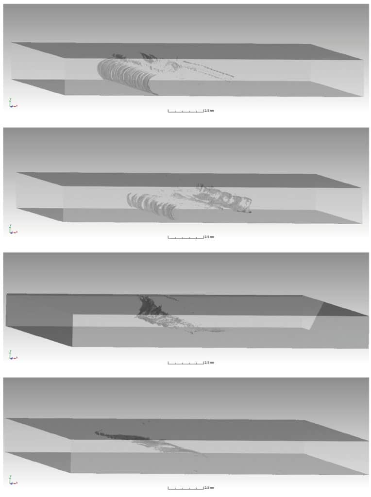
Some of this difficulty in the welding process results in inconsistencies and imperfections in the final weld on a microscopic scale. To identify such deficiencies, Prof Dr Jasiuniene and her team used a combination of X-ray computed tomography and acoustic microscopy to investigate the invisible defects inside welds between different alloys. From the reconstructions of the three dimensional images of the inside of the welds, they could pinpoint exactly where the defects in the weld zones formed. The 3D capabilities of 3D tomography mean all of this can be done in real space and it is possible to resolve defects that would overlap with each other in more standard 2D imaging.

The small size of the nanoparticles, and the defects they can cause, make them very difficult to identify with other non-destructive imaging techniques. The non-destructive nature of the technique also means it is suitable for use on manufactured pieces that require checking and could potentially be done routinely to assess fatigue or accumulating damage.
Self-compacting concrete
Concrete is a mixture of cement and a binder that is usually liquified with water. After the mixture is made, it remains in a gel-type form so it can be poured into moulds and manipulated until it sets as a solid. While it is being poured, the concrete needs to be compacted so that all the air bubbles and voids in the structure are removed to ensure that there are no inhomogeneities in the structure of the concrete and it is as strong as required. This is achieved by using a steel probe to act as a continuous source of vibration while the concrete is being poured.
Self-compacting concrete eliminates the need for continuous vibration and typically has a higher strength for a given water, cement and binder ratio than the traditional form. One way of making self-compacting concrete is to include superplasticisers that help it to be more elastic and flexible than normal concrete. By removing the need for vibration or tamping after pouring and as it also flows very well, self-compacting concrete is much less labour intensive to lay and can be used in areas where it is not possible to compact normal concrete or to create more complex shapes that would not otherwise be achievable.

One type of self-compacting concrete uses steel fibres, much as traditional concrete uses steel bars, to help reinforce the structure. However, it is difficult to know exactly how all of the fibres are aligned after the pouring process, even though the orientation of the fibres is critical in the amount of reinforcement strength the steel fibres will provide along different axes. Visualising the orientation of such fibres, given their small size, is the perfect problem for X-ray computed tomography as it can capture the full size, shape and relative orientation in 3D of the fibres in the concrete.
3D images of the inner structure allows quality evaluation of the different structures non-destructively – making the invisible visible.
Prof Dr Jasiuniene has been applying her expertise in X-ray computed tomography to see how the flow-properties of the concrete influence the eventual orientation of the fibres. Her work has demonstrated that low viscosity concretes show the greatest amount of inhomogeneity in the steel fibre distribution after being poured but also that steel fibres orientate parallel to casting direction after some distance. Meanwhile in high viscosity concrete, fibres are distributed all over the beam more evenly, but they don’t orientate parallel to casting direction as in low viscosity concrete.
Look into the future
X-ray imaging tomography is a highly versatile, non-destructive technique that is widely used in engineering for its ability to recreate 3D images of the insides of many kinds of material. Prof Dr Jasiuniene has been successfully able to use this to better understand the microscopic structure of metal alloys and concretes to help guide manufacturing decisions and reproducibility of the desired mechanical properties of such materials. In the future, she will continue to develop and apply tomography and related methods for the study of not just commonplace concrete, but also for materials used in space engineering and nuclear power plants.
Personal Response
What other material and systems have you been investigating with X-ray imaging tomography?
Investigations of very different types of materials/ structures have been performed: investigation of the structure of 3D scaffolds for bone tissue regeneration; investigation of titanium pin array structure produced using additive manufacturing technology; human teeth for investigation of root canal transportation and centring ability of rotary endodontics instruments; visualisation of inner structure of microchips; investigation of the integrity of concrete in order to assess the harmful consequences of alkali-silica reaction on the composition of the material; investigation of glider longeron, made from CFRP to determine porosity.