Using photodynamic therapy to target circulating tumour cells
Metastasis is life-threatening for patients suffering with cancer. During metastasis, cancerous cells known as circulating tumour cells (CTCs) leave the primary cancer location and enter the bloodstream or lymphatic system. CTCs spread to and accumulate in surrounding tissues and distant organs which then become cancerous. When metastatic cancer develops, the fatality risk increases significantly. In fact, it is estimated that metastatic cancer is responsible for 90% of all cancer-associated deaths. This estimate has changed little in more than 50 years. CTCs were identified in 1869. However studies on these cells were not performed until the late 1900s due to a lack of efficient technology for their detection. Significant advances in technology have led to improvements in detection and isolation techniques such as immunomagnetic separation with antibodies, fibre-optic array scanning technology and microfluidic platforms. Many studies have determined that a high CTC number is associated with poor cancer prognosis. Therefore, it is essential that CTC-specific targeted therapies are developed that eliminate CTCs from the vascular system and improve prognosis. In practice, however, this has been challenging to achieve due to the unpredictable and dynamic nature of CTCs. Dr Choi and his team from Kyung Hee University and Dr Kim from Oncocross, Ltd set out to optimise the ground breaking photodynamic therapy (PDT) to focus on eradicating fluorescent protein-expressing CTCs.

B Scheme of the in vitro fluidic system mimicking blood circulation coupled with laser irradiation.
Specific wavelengths of light activate certain photosensitisers which then convert tissue oxygen into reactive oxygen species and free radicals.
Photodynamic therapy
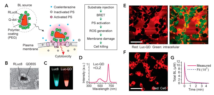
PDT is a treatment method that uses light and specific drugs known as photosensitisers. Specific wavelengths of light activate certain photosensitisers which then convert tissue oxygen into reactive oxygen species (ROS) and free radicals. High levels of ROS can cause oxidative stress, damaging surrounding cellular structures, and can even induce the apoptosis of cancer cells. Although PDT has major biomedical potential, a significant challenge is the delivery of the activation light deep into the tissue. Visible light penetrates a few hundred micrometres in tissue, therefore clinical use of light-based techniques are limited to superficial layers such as the skin and retina. However, there is an interesting solution to this problem – the concept of internal light sources. The implantation of light emitting diodes or fibre-optic light sources are viable, but very invasive for the patient. One attractive alternative to optoelectronic tools is the use of bioluminescence (BL) because the source molecules, such as enzymes, can be delivered with minimal invasion. These bio-luminating molecules can be used in a process called bioluminescence resonance energy transfer (BRET). In BRET, an excited bioluminescent donor (typically a luciferase enzyme) transfers energy to an acceptor. This is a distant-dependent process and the range over which energy transfer can take place is limited to 10 nanometres.
PDT and BRET
To overcome the challenges of using an external light source for PDT, the team investigated the potential of using bioluminescence PDT (BL-PDT), the foundation of which is the BRET phenomenon. Using self-illuminating Renilla luciferase (Rluc8) as the donor protein and photosensitiser chlorine e6 (Ce6) as the acceptor protein, the team showed that BL-PDT can be used to suppress tumour growth in mice for three different cancer cell lines – melanoma, lung cancer and colorectal cancer. Ce6 localises in the cell membrane and organelles such as mitochondria and lysosomes. Activation of Ce6 by energy transfer from Rluc8, results in the generation of ROS, which subsequently induces lipid peroxidation and destruction of the cell membrane. This disrupts intracellular homeostasis and cell death is induced. The team also demonstrated the effectiveness of BL-PDT for cancer cell ablation in the local lymph nodes, reducing metastatic spread and significantly enhancing animal survival. The lymph nodes are located at depths not accessible by external light illumination, therefore BL-PDT could have huge therapeutic potential for targeting metastasis.

Around 60% of GFP-expressing cells were selectively killed when irradiated with 473 nm blue light laser after Rose Bengal treatment.
PDT and FRET
Dr Choi and Dr Kim decided to extend their research by investigating whether this phenomenon could also be adopted in Föster resonance energy transfer (FRET) to specifically target cancer cells. In FRET, light illumination at a specific wavelength excites a donor fluorophore which transfers its excitation energy to a nearby acceptor molecule. In this study, the team used green fluorescent protein (GFP) as the linker of energy transfer between a specific wavelength light source and the photosensitiser Rose Bengal. The team used GFP expressing cancer cells to highlight the function of GFP to target a specific cell population based on PDT. The team had to ensure that the laser excited GFP but had no effect on the photosensitiser to minimise effects on GFP non-expressing cells. Furthermore, the emission spectrum of GFP had to overlap with the absorption spectrum of the photosensitiser to ensure that GFP-expressing cells were selectively killed. As a result, the team chose a 473 nm blue laser. The results showed around 60% of GFP-expressing cells were selectively killed when irradiated with a 473 nm blue light laser after Rose Bengal treatment. However, only 20% of GFP non-expressing cells were killed. This is due to the production of ROS by Rose Bengal upon excitation via energy transfer from excited GFP.

Strikingly, the number of lung metastatic nodules in the treated mice was significantly lower and these mice survived for longer than untreated mice.
Using FRET for CTC Ablation
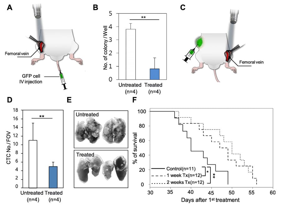
The success of the study using FRET PDT inspired Dr Choi and Dr Kim to target CTC ablation using GFP expressed CTCs and Rose Bengal. The team performed an in vitro study, using a piece of tubing connected to a peristaltic pump to mimic circulation within the blood vessels. GFP-expressing CTCs and GFP-non-expressing CTCs were incubated with Rose Bengal and were passed through the tubing. Results showed that cell damage and death was greater among GFP-expressing CTCs compared to GFP-non-expressing CTCs. An in vivo study was also conducted: by injecting GFP-expressing CTCs and Rose Bengal into mice, a blue laser was then illuminated onto the mouse femoral vein. Results showed that the number of treated CTC colonies dramatically decreased. Additionally, CTC-targeting PDT was performed in mice with GFP-expressing metastatic cells transplanted into their flanks. Again, the number of CTC cells in the irradiated mice was significantly lower than the untreated mice. Strikingly, the number of lung metastatic nodules in the treated mice was significantly lower and these mice survived for longer than untreated mice. Excitingly, these results demonstrate that CTC-targeted therapies aimed at improving cancer prognosis may provide a promising new approach for developing personalised and precision medicine.
Personal Response
How can resonance energy transfer be combined with photodynamic therapy to prevent metastasis?
<> Resonance energy transfer is the key mechanism to eradicate GFP cells selectively. By killing GFP-expressing CTCs, we demonstrated that CTCs might be an effective therapeutic target to significantly delay distant metastasis and ultimately improve patient survival. In addition, it directly suggests CTCs are a core seed to be metastasised into secondary organs. However, these data do not suggest the immediate clinical application of the PDT method examined in this study. Advancements in the field of molecular diagnostics have made it possible to use combinations of fluorescence proteins and photosensitisers or molecular-targeted photosensitisers in diverse biological fields, including not only CTCs-targeted therapy but also cancer stem cell-targeted therapy.