Spiking of urinary biomarker with exercise: A marker of muscle inflammation?
Seeking a sensitive biomarker of inflammation, Emeritus Professor Isao Okayasu of Kiryu University and Kitasato University, Japan and his colleagues have spent over two decades investigating chronic organ inflammation and its links to cancer. They have developed methods to measure prostaglandin E-major urinary metabolite (PGE-MUM) as a surrogate marker, which has opened the door for its investigation in different physiological states. The researchers’ pioneering studies present PGE-MUM as a marker of active inflammation. Now, their most recent study reveals PGE-MUM levels could also reflect muscle inflammation and damage after exercise.
Inflammation is central to disease pathology and helps the body fight foreign invaders and cell or tissue injury. However, this complex process can also occur in the absence of foreign bodies when the body launches a defence against its own cells (an autoimmune response).
Inflammation, orchestrated by a plethora of signalling proteins (cytokines) and mediated by inflammatory cells, including white blood cells, may be acute or chronic. Measurement of signature molecules, called biomarkers, for this process can help monitor disease and evaluate treatment response. Ideally, biomarkers should be simple and easy to measure, while maintaining sensitivity and accurately reflecting the underlying process. One of the most measured biomarkers of inflammation is C-reactive protein (CRP), but its sensitivity and specificity varies in different inflammatory diseases, making it an unreliable marker.
During active inflammation, arachidonic acid is converted by the enzyme – cyclooxygenase 2 (COX-2) – into prostaglandins such as prostaglandin E2. However, this molecule is technically difficult to measure because it is metabolised very quickly. Isao Okayasu, Emeritus Professor of Kiryu University and Kitasato University, Japan has collaborated with a team of researchers to develop methods to measure a proposed surrogate marker of inflammation, prostaglandin E–major urinary metabolite (PGE-MUM) – a by-product of metabolism that is stable and easy to measure.

RIA: radio-immunoassay;
ELISA: enzyme-linked immunosorbent assay;
UC: ulcerative colitis.
Okayasu and collaborators have developed a sensitive radio-immunoassay (RIA) that uses antibodies to measure PGE-MUM (figure 1). The team have conducted years of extensive research to expand the application of PGE-MUM as a biomarker. In their latest research, they investigated whether inflammation of muscles occurs due to physical exercise using urinary PGE-MUM levels. There is now growing evidence that PGE-MUM may be a suitable tool for measuring organ and tissue inflammation in our bodies, opening new avenues for exploration in other diseases.
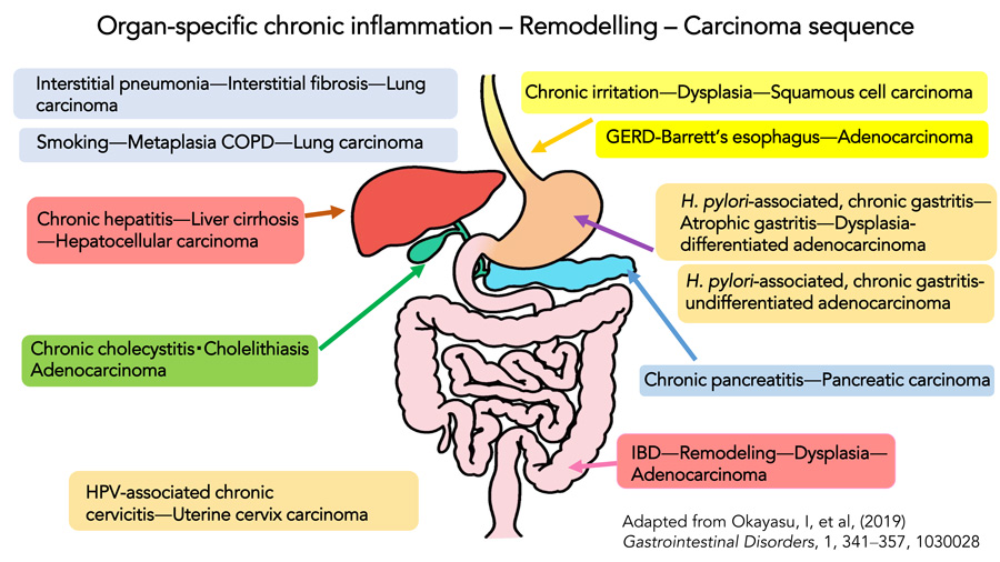
The organ-specific chronic inflammation-carcinoma sequence
The researchers have previously focused on localised chronic inflammation in organs that cause tissue remodelling, and how this can eventually lead to cancer. Such pathology is termed organ-specific chronic inflammation-carcinoma. These cancers may be different to those that stem from non-inflammatory sources. Okayasu proposes that ulcerative colitis (UC) and associated colorectal cancer is an example of organ-specific chronic inflammation-carcinoma. The researchers demonstrated that in humans with UC, certain gut bacteria can enter colon epithelial cells to stimulate inflammatory pathways. Such inflammatory insult and elevated oxidative stress can damage DNA in epithelial cells to cause a mutation in the p53 gene, which is needed to suppress tumours (figure 2).
Inflammation is central to disease pathology and helps the body fight foreign invaders and cell or tissue injury.The researchers developed an animal model of UC known as the DSS colitis model. As mice have similar symptoms to humans, they provide a useful model to study the disease. Importantly, these models revealed that the mucosal epithelium tried to regenerate, but also underwent mucosal erosion and cell death leading to colorectal tumours. Such models provide insight into the pathology of colorectal tumours in UC and offer avenues to prevent and treat the disease. Treatments such as anti-inflammatories help arrest the chronic inflammation that is fundamental in UC pathophysiology.
Early studies of prostaglandin E–major urinary metabolite
For any marker or measurement, it is important to first understand how it varies across the population, especially with age and gender. The researchers’ previous studies indicated that PGE-MUM levels were higher in biological males than in biological females. These levels decreased with age in males but increased with age in females. Several reasons for this difference are suggested, including hormonal changes in both sexes. However, Okayasu’s recent work showed that the influence of sex hormones on PGE-MUM is significant but small. Interestingly, increased levels of PGE-MUM, regardless of age, were found in smokers. The researchers suggested that this reflects inflammation and injury in the lungs caused by smoking. This biomarker was elevated only in current smokers; conversely, CRP is increased in both current and ex-smokers. The team propose that, in conjunction with clinical assessment, PGE-MUM could be used to screen and determine inflammation in the body, offering an alternative marker to CRP.

UC presents as ulceration of the mucosal lining in the large intestine. Colonoscopy allows examination of the large intensive using a camera to monitor and assess mucosal healing. However, this is an invasive and often uncomfortable procedure. Non-invasive tests for biomarkers of UC are therefore needed. In particular, sensitive and specific UC biomarkers are lacking, since CRP has questionable sensitivity.
In their landmark study in 2014, the team showed that PGE-MUM offers a sensitive test and better reflects UC activity compared to CRP. The test only requires urine samples and is simple, cost-effective, and quick to carry out. The researchers propose that PGE-MUM can be used to monitor patients in remission to check whether the inflammation indicative of active disease is increasing. The researchers also reported that PGE-MUM detects colonoscopic remission more accurately than CRP. However, Okayasu emphasises that additional studies are needed to check if PGE-MUM levels can predict relapse and active disease over time.
Physical exercise and muscle inflammation
Mild and extreme physical exercise can stimulate an inflammatory response by increasing cytokines. This inflammation can cause muscle damage. In fact, studies on muscle biopsies after mild exercise show that mRNA for COX2 is elevated following activity. Theoretically, this could lead to increased PGE2 and PGE-MUM. Okayasu’s groundbreaking work builds a case for PGE-MUM as a useful marker of inflammation in different physiological settings. Expanding on this research, the research team are now exploring the effects of physical exercise on PGE-MUM levels.
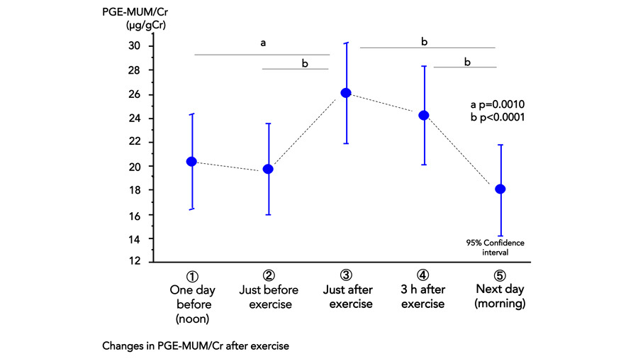
The researchers describe how PGE-MUM, standardised by correcting for the concentration of urinary creatinine (an indicator of muscle mass and performance), was measured in teenage boys at time points before and after physical activity. The team conducted two separate sub-studies to assess different intensities of exercise. In the first study, the participants played the sport soccer, to represent mild exercise. The second study was conducted over three days and participants underwent repeated exercise, such as running. PGE-MUM levels and protein levels, including urinary L-type fatty acid-binding protein (L-FABP), were measured and compared with PGE-MUM levels (figure 3).

The researchers demonstrated that both types of exercise result in elevated PGE-MUM levels. In mild exercise, PGE-MUM levels increased significantly after exercise by peaking first and then decreasing over the next few hours and eventually returning to pre-exercise levels on day two. In the study of repeated exercise, PGE-MUM levels peaked on the second day after the participants ran uphill and downhill. The levels also increased following long-distance running on a flat surface, but these levels were not as high as those following the uphill and downhill run. The researchers suggest that this could be due to the different nature and severity of exercise, including the muscle fibres required.
There is now growing evidence that PGE-MUM may be a suitable tool for measuring organ and tissue inflammation in our bodies.Incidentally, the team noted an increase in L-FABP and total urinary protein levels as markers of renal function after exercise, suggesting temporary kidney stress post exercise. With the strong correlations between PGE-MUM and urinary creatinine, the team speculate that the increased PGE-MUM levels after exercise reflect muscle inflammation due to exercise, although they acknowledge that this could be related to the kidney. The researchers argue that their simple, non-invasive test could be carried out before and after exercise to serve as a specific marker of muscle damage and inflammation.
The research of Okayasu and colleagues – developing a method to measure PGE-MUM – provides promising data. It could be a useful and cost-effective tool to measure inflammatory status in human bodies. Their study serves as a starting point for understanding more about the role of PGE-MUM to assess various physiological states as well as diseases.

Okayasu maintains that there is scope for further studies to confirm the findings and expand the understanding of this metabolite. Technological advances in detection methods will enable rapid analysis of easily accessible urine samples. This will also invite future large-scale studies to assess PGE-MUM as a marker of inflammation. With their dedicated efforts in exploring PGE-MUM under conditions of inflammation, Okayasu and colleagues hope to unlock and fully elucidate the potential of this biomarker.
Personal Response
Do you have plans to extend the PGE-MUM work by studying other conditions where inflammation is central?First, we are investigating whether PGE-MUM might be a possible marker for muscle inflammation due to physical exercise, in practical terms. Second, we continue to investigate which factors differ with age and gender, in healthy subjects. Third, we should clarify what causes the individual difference in PGE-MUM because it is rarely large without any reason.