Electrical eye activity reveals early sight damage in alcohol addicts
People addicted to alcohol often suffer loss of vision through damage to the light receptors and nerves in the eyes. Scientists have previously shown interest in measuring the electric currents in the brain related to vision, but the information gathered has not yet made an impact on preventing further sight loss. Professor Haolin Zhang at the Beijing University of Technology, and Ms Xin Xie at Peking University Third Hospital, China, have been looking into a more holistic way of assessing electric eye activity and it seems to be of great clinical potential.
Vision is a highly specialised sense that allows humans to perceive and explore their surroundings by interpreting light stimuli reflected from their environment. The visual pathway is the route by which light stimuli are transferred through the eyes to the occipital cortex of the brain, the area responsible for the sense of vision. The retina is a light-sensitive layer of tissue that lines the inside of the back of the eye. It consists of two main types of light receptors: rods and cones. Rods are cylindrical cells sensitive to dim light while cones are conical cells active in brighter light and responsible for colour vision. With the help of these cells, the retina converts light energy into electric signals which are then transferred through the optic nerves to the brain’s visual centre in the occipital lobe (the surface area of the lower-back side of the brain). Other cells of the retina involved in the process are the amacrine cells, which are nerve-connecting cells responsible for high vision sensitivity in dim light environments, and ganglion cells, the nerve cells that lie between the light receptors and the area of formation of the optic nerve, connecting the two together.
How alcohol affects vision
Chronic alcohol consumption has been linked to impaired vision. More specifically, alcohol can damage various parts of the visual pathway, including the nerve that carries the visual information to the brain (a condition known as optic neuropathy) and the retina. Alcohol has also been shown to damage the little blood vessels that supply the retina with oxygen and nutrients. Its toxic effects could also be attributed to the fact that alcohol reduces the activity of gamma-aminobutyric acid (GABA), an important neurotransmitter (a signalling molecule secreted by a nerve cell to affect another nerve cell across the synapse that connects them), which causes dysfunction of the retina cells.

The effects of chronic alcohol consumption are initially reversible, especially since the disorders are functional and mainly consist of decreased sensitivity of the light receptor cells and optic nerve without any structural changes. However, if the alcohol abuse continues it can cause structural damage to the optic nerve and retina, leading to permanent vision decline and occasionally to complete vision loss. Professor Haolin Zhang at the Beijing University of Technology and Ms Xin Xie at Peking University Third Hospital in China aim to develop a diagnostic tool that would allow timely intervention to prevent temporary vision damage in alcoholics from becoming permanent.
Alcohol can damage various parts of the visual pathway, including the optic nerve and the retina.Electric currents tell stories
The electric currents into which the light stimuli are converted on the retina can be detected and measured by a diagnostic tool called an electroretinogram (ERG), which is considered to be an objective and accurate way of assessing retinal function. The detection of electric signals in this way is not unlike electroencephalography (EEG), which records the brain’s electric activity through electrodes attached to the scalp. Since the position of the eye makes it easier to access than the brain, the researchers believe that the data provided by the ERG might be more accurate than the information acquired using EEG. In their study, they used both methods as a more comprehensive approach for early detection of alcohol-induced damage to the retina. Specifically, they used full-field electroretinography (ffERG), a type of electroretinogram that better quantifies the retina lesions, and electrophysiology tests for visually generated signals called pattern-reversal visual evoked potentials (PR-VEPs).
An eye-opening experiment
The study conducted by Zhang and his team involved 11 alcoholics and 14 non-alcoholic individuals as a control group matched for age, gender, and smoking habits. The participants underwent testing with both ffERG and PR-VEPs between September 2017 and March 2022 at the Department of Ophthalmology of Peking University Third Hospital. The inclusion criteria were more than two years of addiction to wine, drinking 100ml of 40–50% alcohol-content drinks at least three times a week, and the absence of any other non-alcohol related eye disease.
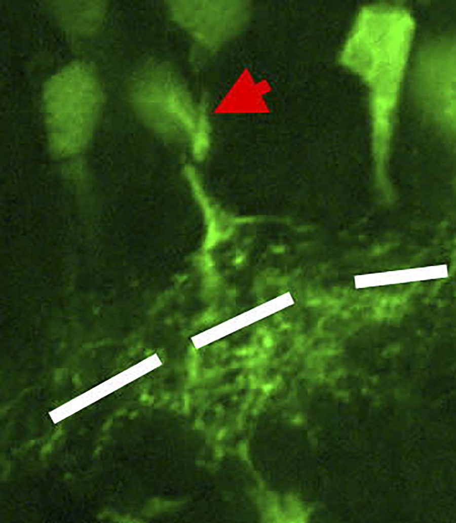
To investigate the toxicity of alcohol on rods, cones, and other retina cells, the team ran ffERG tests both in dim and bright light (scotopic ffERG and photopic ffERG respectively). They also took digital photographs of the eye fundus – the inside of the back of the eye where the retina and the entry of the optic nerve can be seen. For the tests and data processing they used a device called a Ganzfeld electrophysiology system.
After acquiring all the information available, the team identified 29 electrophysiological parameters for ffERG and ten for PR-VEP. They then analysed the information using multivariate statistical analyses (MVA), including the Permutational Multivariate Analysis of Variance (PERMANOVA) test. The PERMANOVA test is helpful when scientists want to compare multiple parameters in two groups of objects that might or might not be correlated.
Alcohol effects on circulation and connecting cells
From their results, the researchers discovered some significant changes in the retina of the participants with alcohol addiction, in particular that the electric currents were less potent when alcohol addicts were tested in dim light. Since there were no differences found in rod function between the two groups, the team concluded that the findings could indicate impaired local blood circulation in the individuals with alcohol addiction and also an amacrine cell dysfunction that negatively affected dim-light vision.
Their methods could be extremely valuable in detecting early alcoholic vision decline, especially since alcoholics do not often perceive the functional damage to their eyes themselves until they have more serious symptoms.The PR-VEPs results mainly provided information on the function of the optic nerves and the ganglion cells. The optic nerves of alcohol addicts were less able to conduct the electrical impulses they received, and impaired function of the ganglion cells was also seen, consisting of lower response rates to stimuli in individuals with alcohol addiction compared to the control group.
The fundus photographs of everyone in the control group and most of the people in the alcohol addicts’ group were normal, suggesting that functional vision changes can occur without any obvious damage of the retina.

An accurate vision evaluation tool?
Zhang and colleagues’ study takes a more holistic approach to the eye and vision-related brain electric activities. Their measurement of 39 different parameters of the electric currents detected, and use of multivariate non-parametric statistical analysis, resulted in more accurate and comprehensive interpretation of the data than other approaches. The changes they discovered in the eyes of the alcohol addicts were sensitive and reflective of the level of alcohol damage; therefore, their methods could be extremely valuable in detecting early alcoholic vision decline, especially since alcoholics do not often perceive the functional damage to their eyes themselves until they have more serious symptoms.
This novel testing strategy can provide a non-invasive and affordable tool to accurately evaluate visual pathway conditions in alcohol addicts and help clinicians to make early interventions before any damage becomes permanent.
Personal Response
Dr Zhang, could you tell us more about how you identified the electrophysiological parameters? What were the criteria for using them in your study?The inspiration came from where I previously dealt with transcriptome sequencing data in genetics research. The electrophysiological parameters and the gene expression data are intrinsically similar in that they are both vast in amount, have extreme values, and do not follow the normal distribution. Then I narrowed it down, in the same way I identified the most differentially expressed genes.
From 1865, when Swedish physiologist Holmgren first recorded the ERG from frog retina, visual electrophysiology has made significant progress, accompanied by the designation of relevant criteria. We used the latest criteria designated by the International Society for Clinical Electrophysiology of Vision.